Scanning electron microscope images of beaded Sepharose CL-6B with 100
Par un écrivain mystérieux
Last updated 16 juin 2024


Causes of Industrial Protein A Column Degradation, Explored Using Raman Spectroscopy

Preparation of DAMDPA-activated Sepharose CL-6B by periodate oxidative
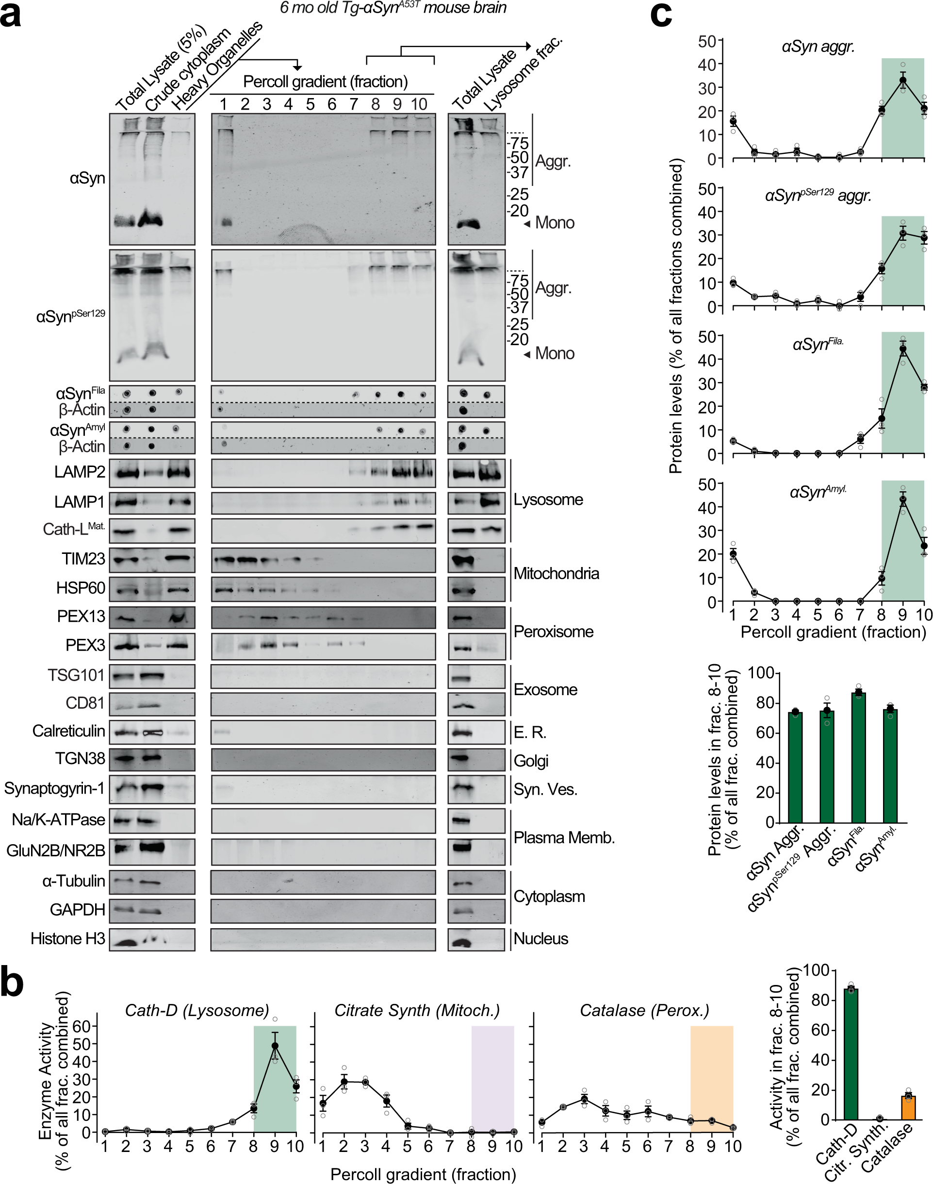
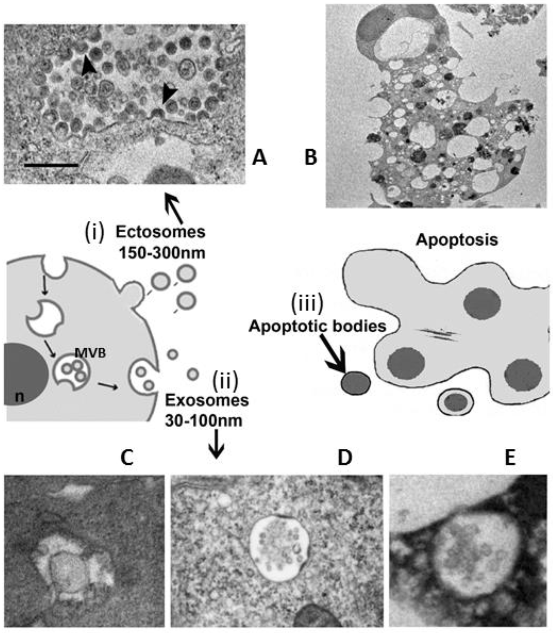
Lysosomal exocytosis releases pathogenic α-synuclein species from neurons in synucleinopathy models
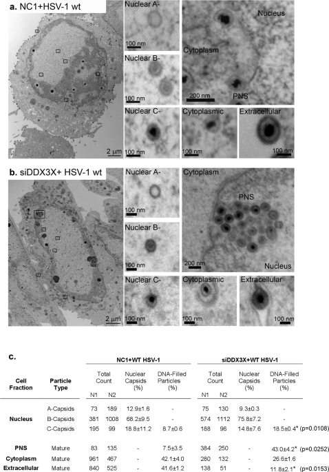
RNA helicase DDX3X modulates herpes simplex virus 1 nuclear egress

Scanning electron microscope images of beaded Sepharose CL-6B with 100
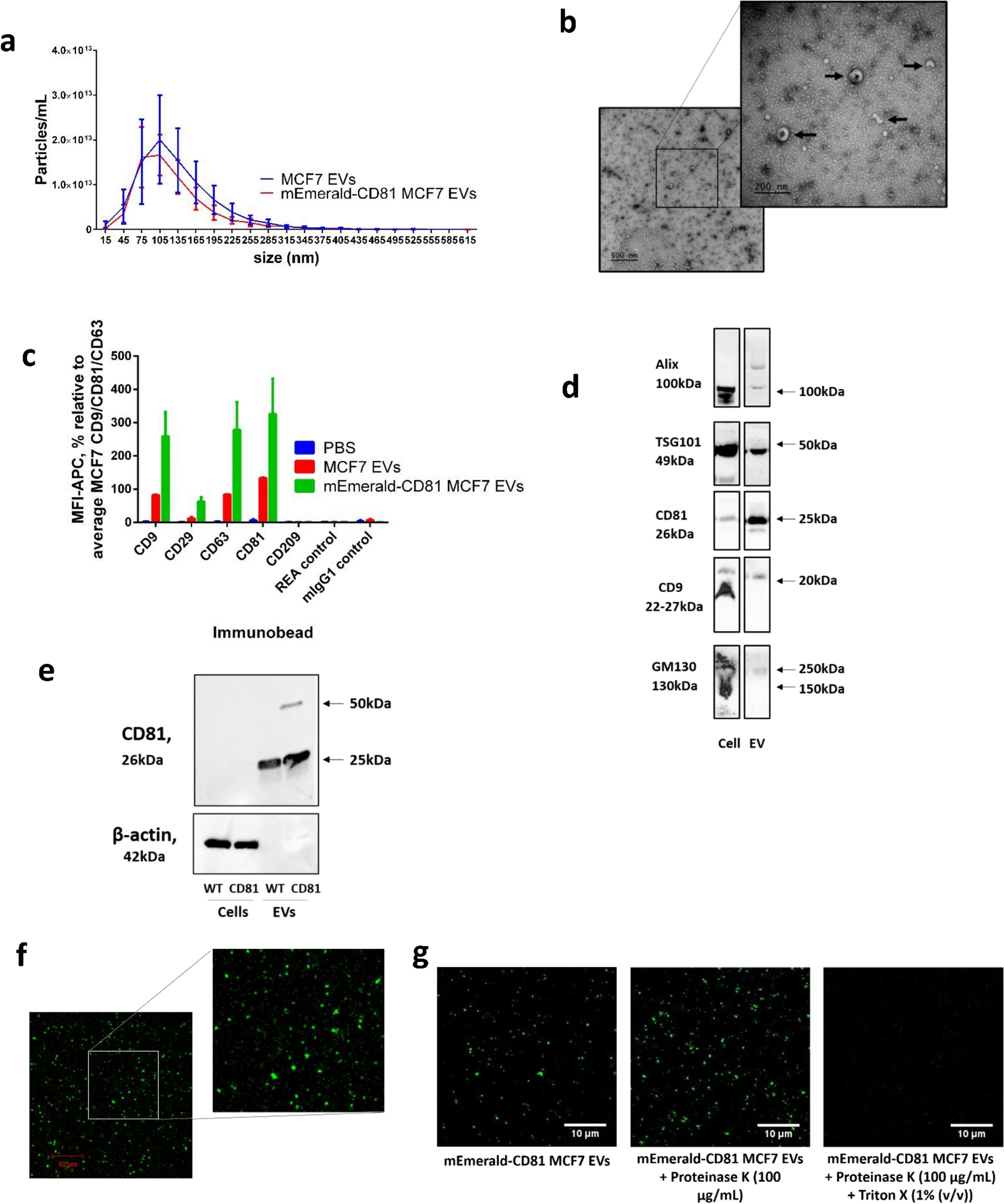
Confocal microscopy analysis reveals that only a small proportion of extracellular vesicles are successfully labelled with commonly utilised staining methods
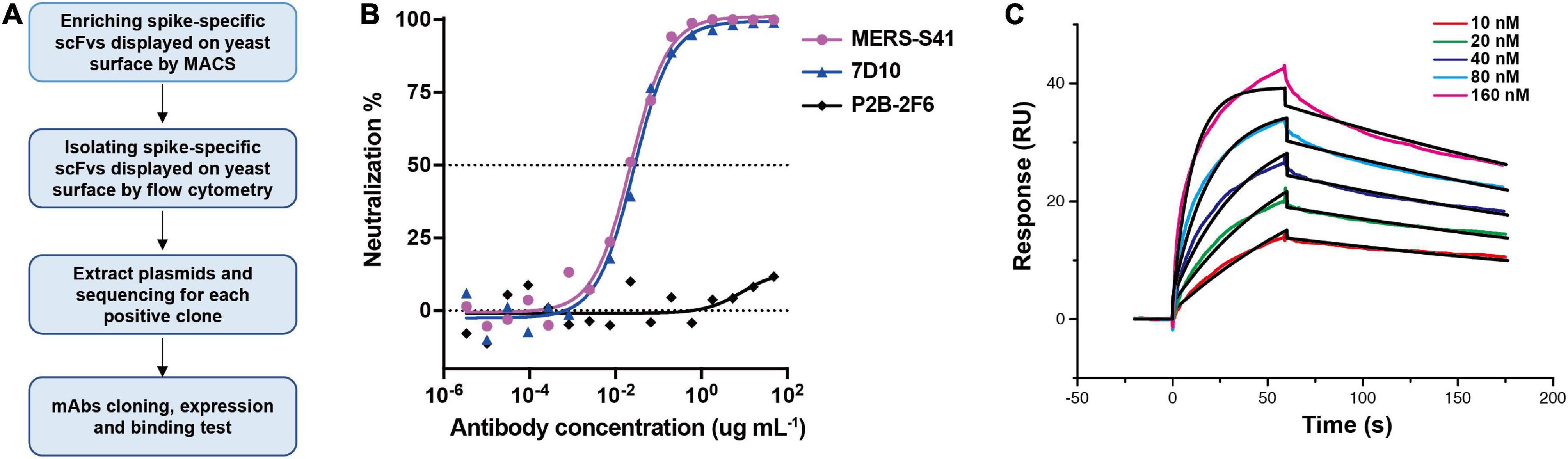
Frontiers Cryoelectron microscopy structures of a human neutralizing antibody bound to MERS-CoV spike glycoprotein

IJMS, Free Full-Text
Biology, Free Full-Text

Mechanical characterisation of agarose-based chromatography resins for biopharmaceutical manufacture - ScienceDirect
Recommandé pour vous
 Carson Microflip 100-250x High Power Pocket Microscope14 Jul 2023
 Carson portable microscopes - Cloudy Days & Microscopes - Cloudy Nights14 Jul 2023
 Micro-Science 92002 100/250/500X HD New Generation Microscope, Yellow/Red : Toys & Games14 Jul 2023
 Microscope Advanced - Binocular — Eisco Labs14 Jul 2023
 AmScope 40X-2500X USB-C Rechargeable Student Binocular Compound Microscope w/3D Two-Layer Mechanical Stage, Achromatic Objective - AliExpress14 Jul 2023
 Objective Working Distance 0.25mm 100X Infinity Plan Achromatic Objective BM0253383114 Jul 2023
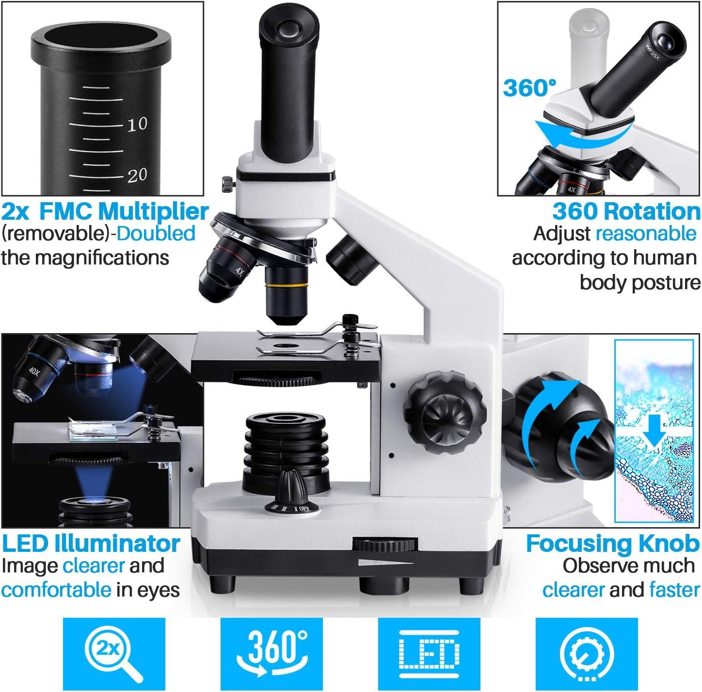 MAXLAPTER Microscope for Adults Kids Students 100-2000x Powerful Biological Educational Microscopes with Operation Accessories (10p), Slides Set (15p), Phone Adapter, Wire Shutter & Backpack Wr855(white)14 Jul 2023
 alga HD Microscope, 100/250/500x - buy at Galaxus14 Jul 2023
 Microscope for Kids,40X-2000X Compound Microscope Mobile Phone Adapter with Microscope Slide Microscope kit for Home School Laboratories for Children14 Jul 2023
 Carson MicroFlip MP-250 100-250x Pocket Microscope14 Jul 2023
Tu pourrais aussi aimer
 This NES Classic Mini knockoff looks dangerously close to the real thing – NintendoSoup14 Jul 2023
 Philips Hue - Plafonnier connecté STILL - 32W - Blanc - White Ambiance - Télécommande Hue incluse - Lampe connectée - Rue du Commerce14 Jul 2023
 Lot 3 pinceaux aquarelle ronds Raphaël petit gris série 83514 Jul 2023
 BB&Co - Matelas à langer nomade en coton gaufré nuage par BB&Co14 Jul 2023
 Infantino coffret éveil bébé - Infantino14 Jul 2023
 Bandeau Affichage Panneau Lumineux - 420 LED Rouge - LABEL sa - occasion14 Jul 2023
 Le proche infrarouge14 Jul 2023
- Anneau lumineux DEL de 6 po Mobifoto Mobilite avec trépied de14 Jul 2023
 113 photos et images de Prise Réseau - Getty Images14 Jul 2023
 Pack de 4 Cartouches d'encre compatible avec HP 963 XL 963XL pour HP Officejet PRO - Cartouche d'encre - Achat & prix14 Jul 2023
