Electron microscopy of stretch-grown axons. Scanning electron
Par un écrivain mystérieux
Last updated 20 juin 2024

Download scientific diagram | Electron microscopy of stretch-grown axons. Scanning electron micrographs illustrating a small fascicle composed of axons 100-250 nm in diameter (A, B). Fasciculation of axons occurs during the elongation process as smaller bundles and individual axons coalesce and adhere to one another, forming larger bundles similar to the one depicted here. Transmission electron micrograph of cross sections near the center of axon fascicles in nonstretch conditions ( C) and axons stretched to a length of 5 cm in 14 d (D), showing no change in axon cytoskeletal structures. Scale bars: A, 10 m; B, 1 m; C, D, 500 nm. from publication: Extreme Stretch Growth of Integrated Axons | Large animals can undergo enormous growth during development, suggesting that axons in nerves and white matter tracts rapidly expand as well. Because integrated axons have no growth cones to extend from, it has been postulated that mechanical forces may stimulate axon | Axons, Growth Cones and Afferent Neurons | ResearchGate, the professional network for scientists.
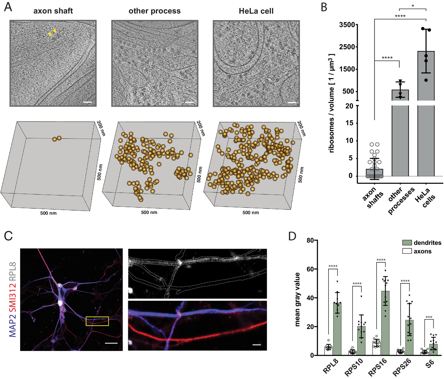
Electron cryo-tomography reveals the subcellular architecture of growing axons in human brain organoids
Three dimensional electron microscopy reveals changing axonal and myelin morphology along normal and partially injured optic nerves
Micromachines, Free Full-Text

Magnetically-actuated microposts stimulate axon growth - ScienceDirect

Extremely Low Forces Induce Extreme Axon Growth

Extreme Stretch Growth of Integrated Axons

Axonal plasticity in response to active forces generated through magnetic nano-pulling - ScienceDirect
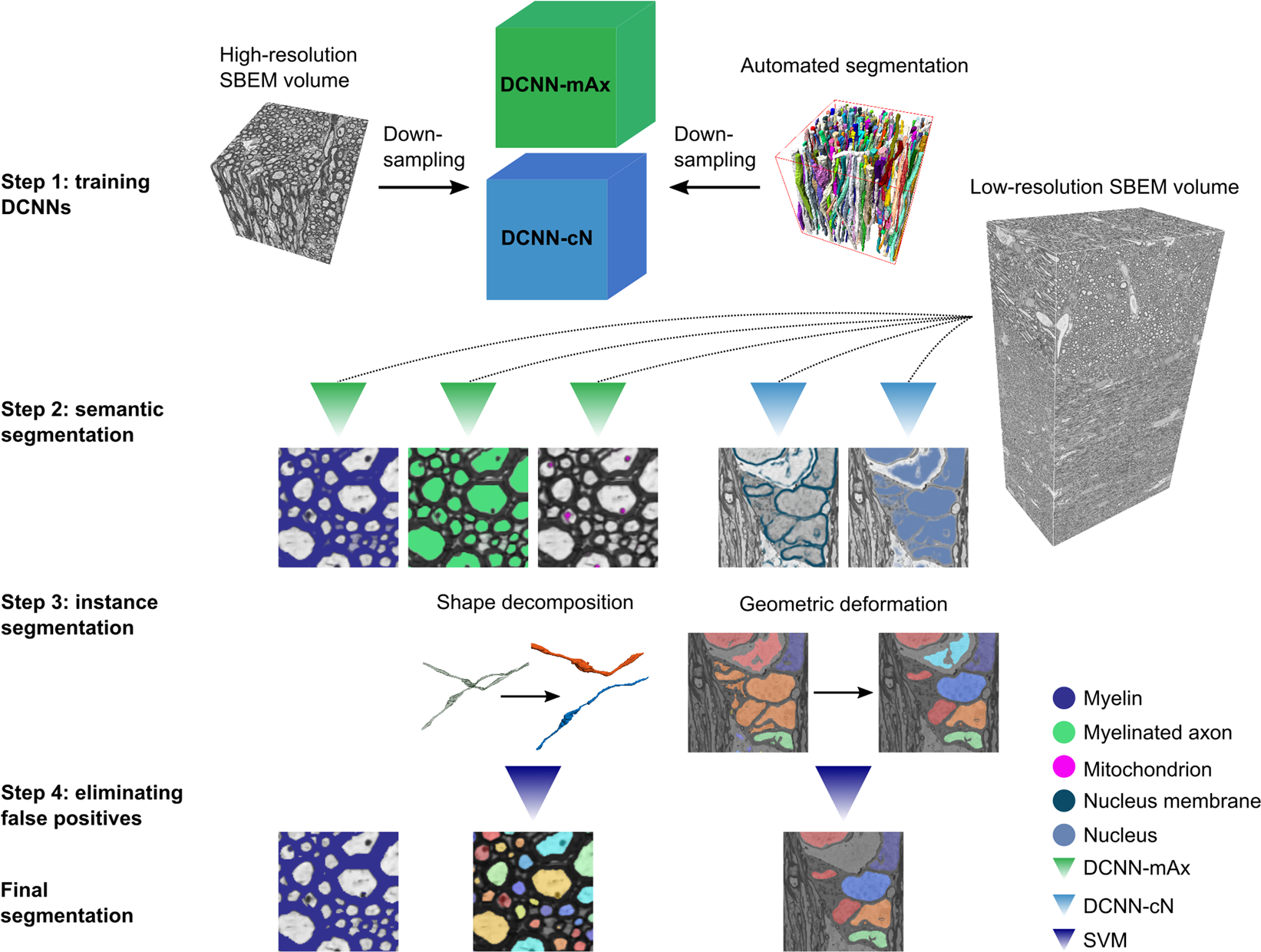
DeepACSON automated segmentation of white matter in 3D electron microscopy

Transmission Electron Microscopy and Morphometry of the CNS White Matter

Incidental Ultrastructural Findings in the Sural Nerve and Dorsal Root Ganglion of Aged Control Sprague Dawley Rats in a Nonclinical Carcinogenicity Study - William A. Meier, Michael J. Linn, Wendell P. Davis

Cells, Free Full-Text
Recommandé pour vous
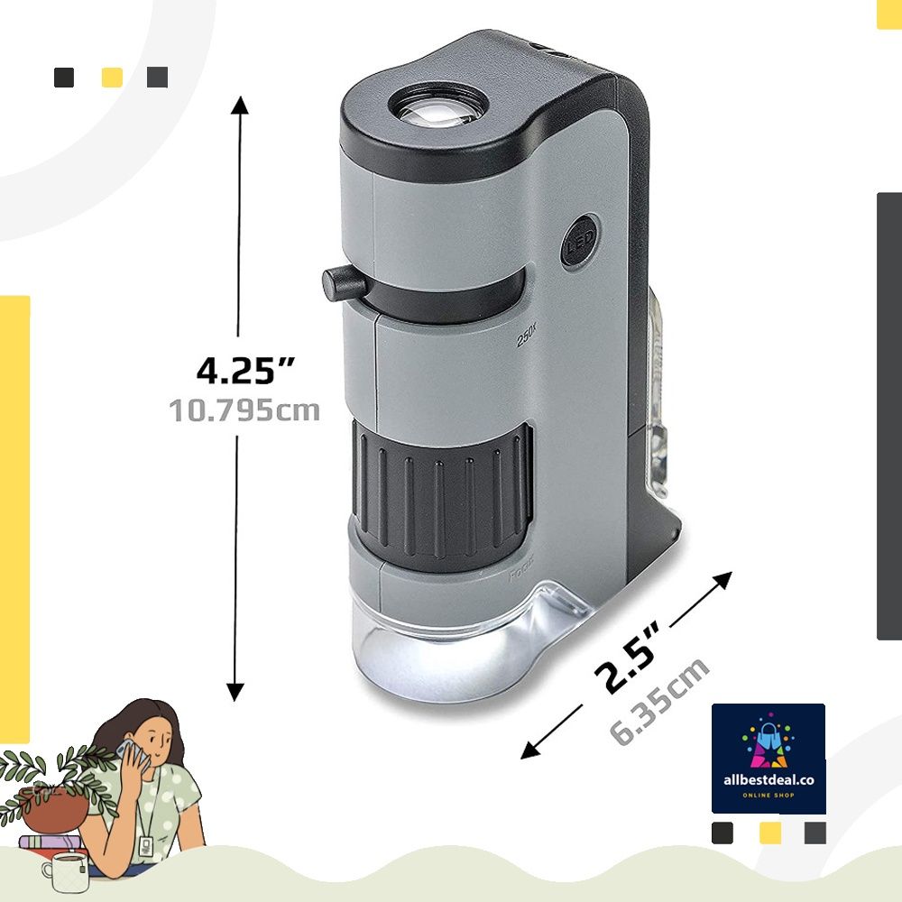
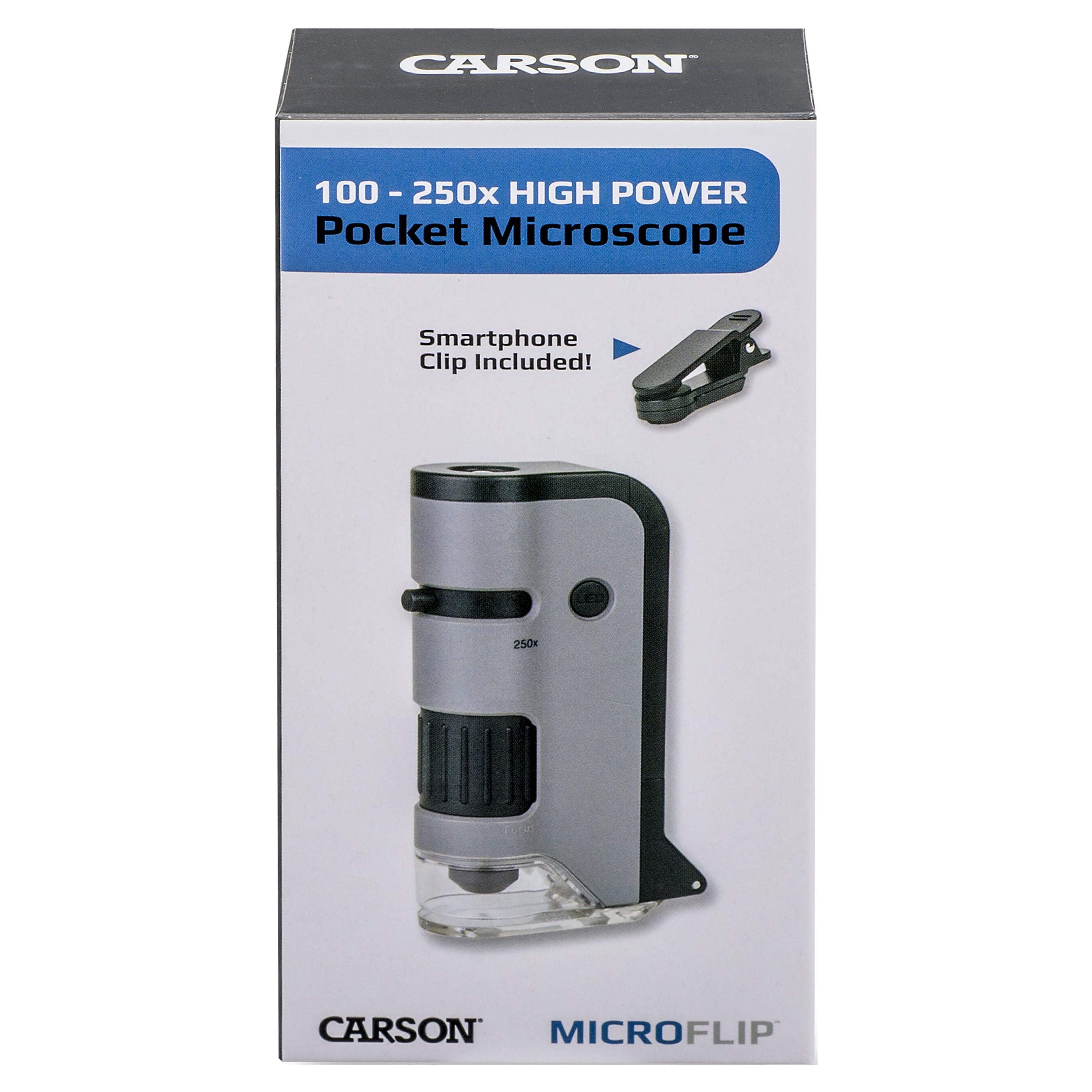 Carson MicroFlip 100x-250x LED Lighted Pocket Microscope with Flip Down Slide Base, Smartphone Adapter Clip, and UV Flash14 Jul 2023
 Carson Microflip 100-250x High Power Pocket Microscope14 Jul 2023
 Carson MicroFlip 100x-250x Microscope de Poche LED et UV avec Base coulissante Rabattable et Clip de numérisation pour Smartphone (MP-250 ou MP-250MU)14 Jul 2023
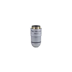 Objective Working Distance 0.25mm 100X Infinity Plan Achromatic Objective BM0253383114 Jul 2023
 SEM (Scanning Electron Microscope) microphotographs of manganese14 Jul 2023
 Microscope For Adults Kids Students 100-2000x Magnification Powerful Biological Educational Microscopes14 Jul 2023
 MAXLAPTER Microscope for Adults Kids Students 100-2000x Powerful Biological Educational Microscopes with Operation Accessories (10p), Slides Set (15p), Phone Adapter, Wire Shutter & Backpack Wr855(white)14 Jul 2023
 Microscope Glass Cover Slides Coverslips,18 mm x 18mm (1pkt of 100) at Rs 199.00, Frosted Glass Slide, Lab Glass Slides, माइक्रोस्कोपिक ग्लास स्लाइड - Labpro International, Ambala14 Jul 2023
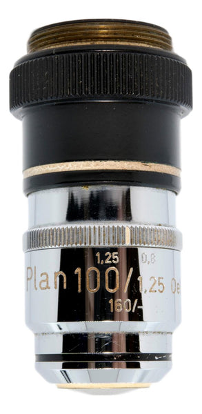 Zeiss 100x Plan Correction-Collar Oil Objective – Microscope Central14 Jul 2023
- instock~ Carson MicroFlip 100x-250x LED and UV Lighted Pocket Microscope with Flip Down Slide Base and Smartphone Digiscoping Clip Bundle Included with 24 Prepared Insect and Animal Slides (MP-250BUN), Health & Nutrition14 Jul 2023
Tu pourrais aussi aimer
 Calendrier Perpétuel à imprimer14 Jul 2023
 Chargeur portable 10000mAh pour Apple AirPods Pro 2 Huawei P1014 Jul 2023
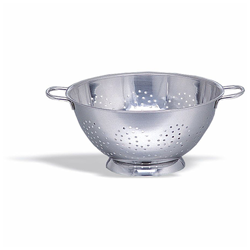 Passoire-Passoire Inox Sur Pied 30 Cm Pujadas - Promark14 Jul 2023
 rotatif Dremel Brosses à fil Roues de polissage Brosse de roue de fil d'acier14 Jul 2023
 PASSOIRE VACUVIN Cocktail Strainer14 Jul 2023
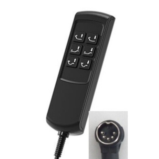 Télécommande TOPLINE 6 Boutons prise DIN coudée 5 broches14 Jul 2023
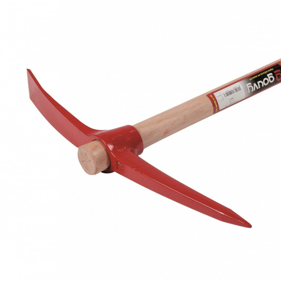 Pioche ronde 2,5kg MANCHE FIBRE (90cm) - Muller - 05408114 Jul 2023
 Adultes Cow-Boy Double Étui Avec Guns Pistolet Revolver Far West Six Tir Crème14 Jul 2023
 10 accessoires déco seconde main à absolument avoir chez soi - M614 Jul 2023
 Petit électroménager - My Home Passion14 Jul 2023