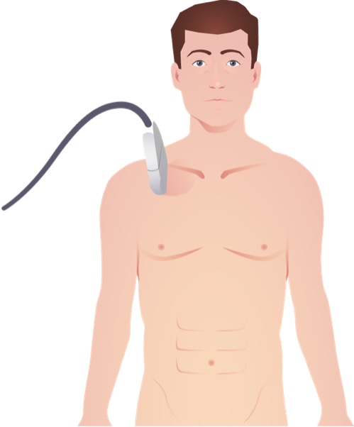
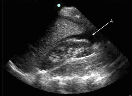
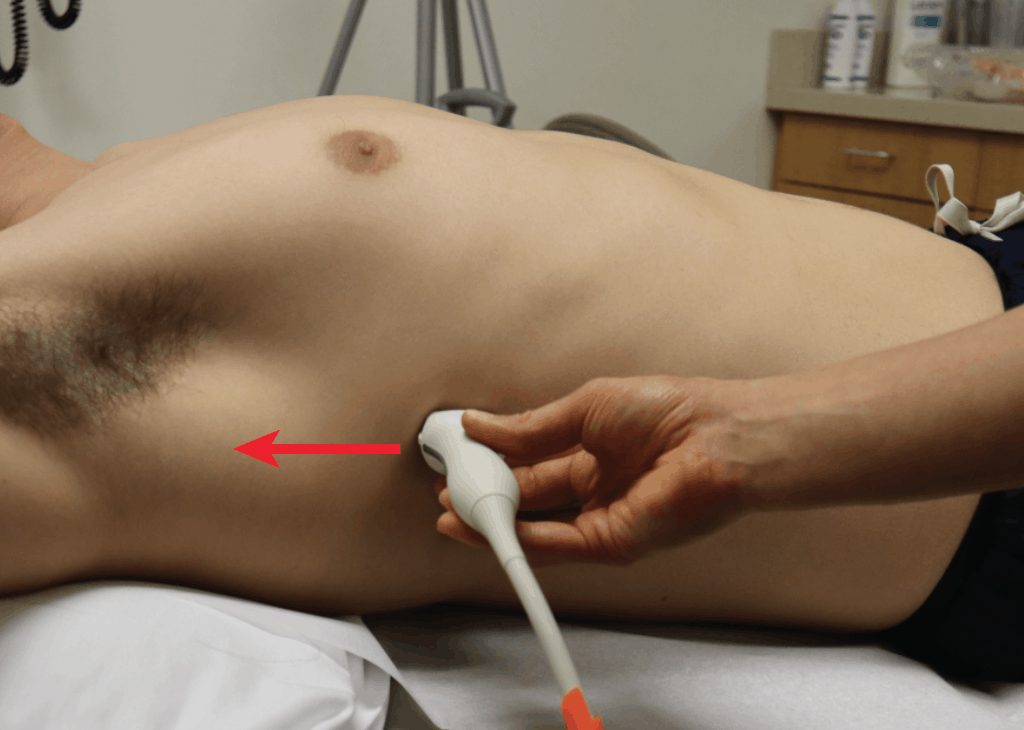
Probe position and normal images obtained during E-FAST examination.
Par un écrivain mystérieux
Last updated 01 juin 2024

Download scientific diagram | Probe position and normal images obtained during E-FAST examination. (A) Right upper quadrant view demonstrating interface between liver and kidney (Morison's pouch). (B) Left upper quadrant view demonstrating spleen and kidney interface. (C) Left transverse view of the bladder. (D) Subcostal or subxiphoid view using the liver as a window to view the heart. (E) Anterior lung view. (F) Anterior lung view with US set to motion mode: this depicts a 1-dimensional view (marked on the top of the screen) as it changes over time (marked on the bottom of the screen); straight lines represent static soft tissue above the granular pattern representing the sliding of the pleura back and forth over time. E-FAST, Extended Focused Assessment with Sonography in Trauma examination. Used by permission from Introduction to Bedside Ultrasound, Vol 1, Dawson M, Mallin M, eds. Lexington, KY: Emergency Ultrasound Solutions; 2012: chap 1. from publication: Point-of-Care Ultrasound in Established Settings | The original and most widely accepted applications for point-of-care ultrasound (POCUS) are in the settings of trauma, shock, and bedside procedures. Trauma was the original setting for the introduction of POCUS and has been standardized under the four-plus view examination | Point-of-Care Systems, Ultrasound and Ultrasonography | ResearchGate, the professional network for scientists.
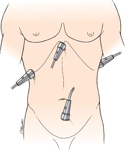
Focused Assessment with Sonography for Trauma (FAST)
Ultrasound in Trauma Critical Care
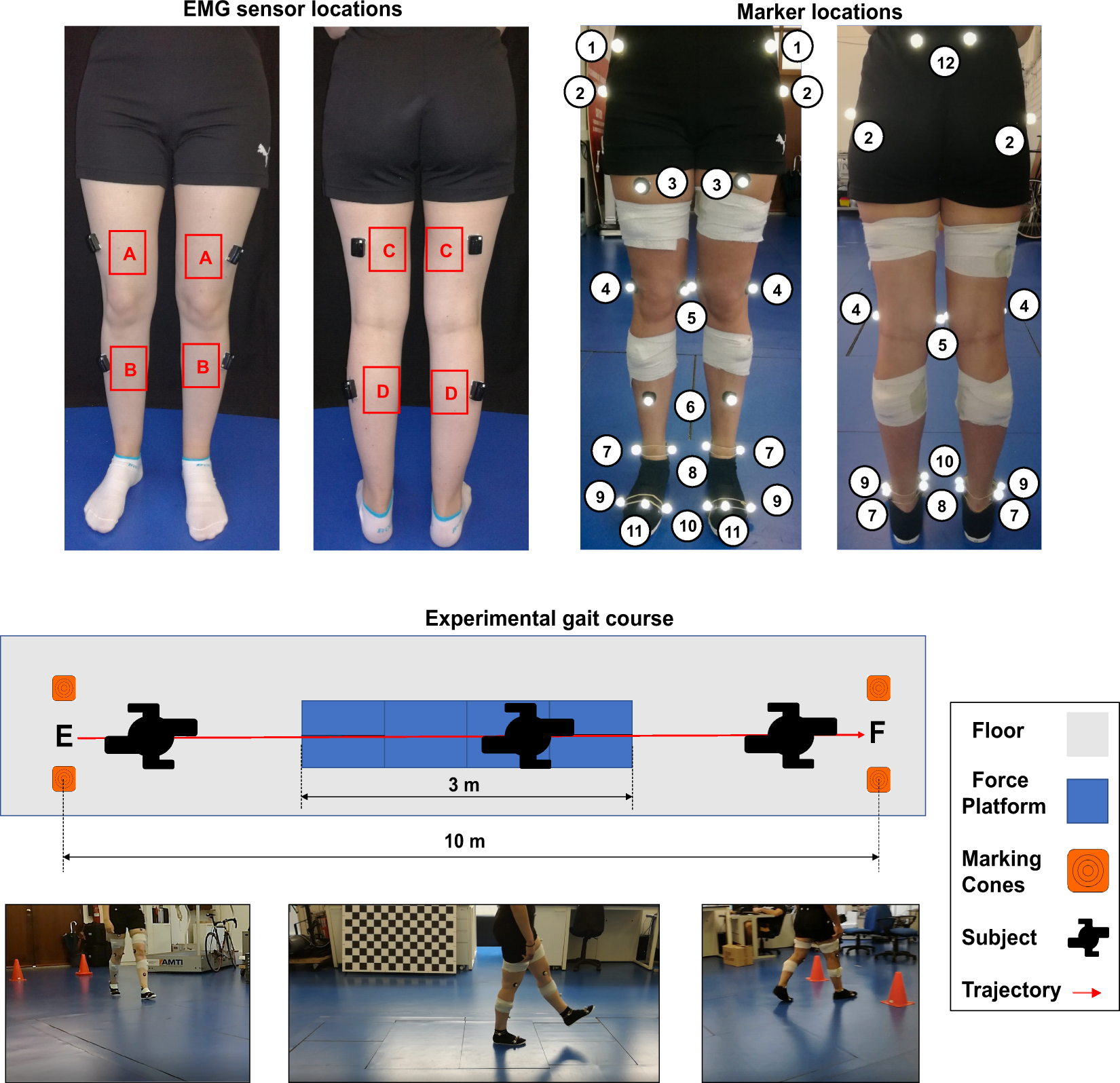
Lower limb kinematic, kinetic, and EMG data from young healthy humans during walking at controlled speeds
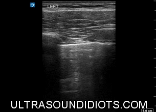
Ultrasound Idiots — Trauma / EFAST

How To Do E-FAST Examination - Critical Care Medicine - Merck Manuals Professional Edition
eFAST Ultrasound Exam Made Easy: Step-By-Step Guide - POCUS 101
Bedside Ultrasound For Surgeons

Obstetric Dopplers – ULTRASOUNDPAEDIA
EMERGENCY ULTRASOUND
Recommandé pour vous
 Use of eFAST in Patients with Injury to the Thorax or Abdomen14 Jul 2023
 Trauma – RMG Ultrasound14 Jul 2023
 Ali on X: eFAST views and questions. #FOAMed #FOAMus #eFAST #POCUS @PsmmcER FF=free fluid. / X14 Jul 2023
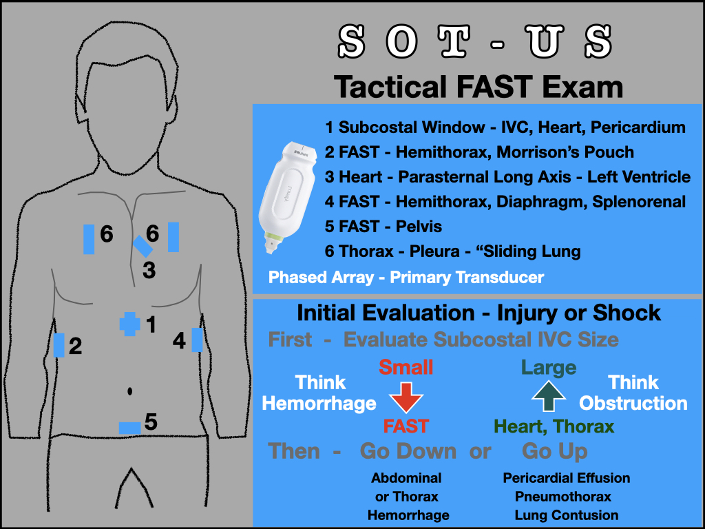 Tactical POCUS14 Jul 2023
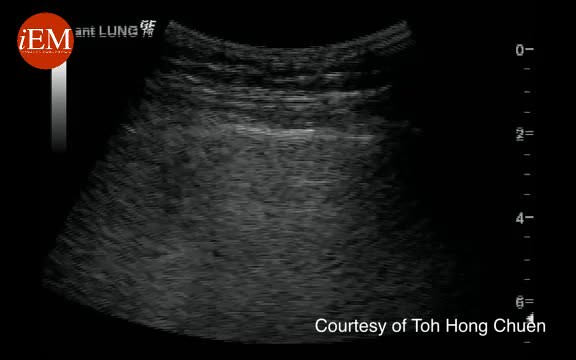 eFAST, Extended Focused Assessment using Sonography in Trauma14 Jul 2023
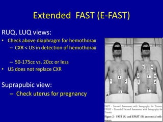 Fast Scan14 Jul 2023
 What is e-FAST?14 Jul 2023
 eFAST – International Emergency Medicine Education Project14 Jul 2023
 Fast letter e logo icon design Royalty Free Vector Image14 Jul 2023
 🔍 FIRST LOOK: Fast-charging in Formula E ⚡️14 Jul 2023
Tu pourrais aussi aimer
 Epson Expression Home XP-2200 - imprimante multifonctions - couleur14 Jul 2023
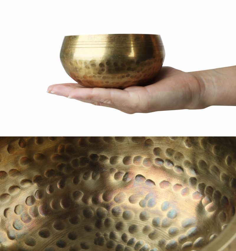 Kit de bol chantant tibétain - Bleu14 Jul 2023
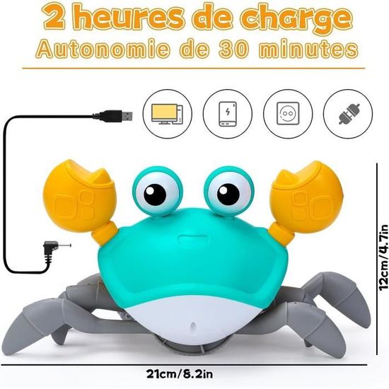 Crabe Qui Marche Bébé - Jouet Bebe 1 2 Ans avec Lumière LED et Musique Jouets Musicaux Evite Automatiquement Les Obstacles - Cdiscount Jeux - Jouets14 Jul 2023
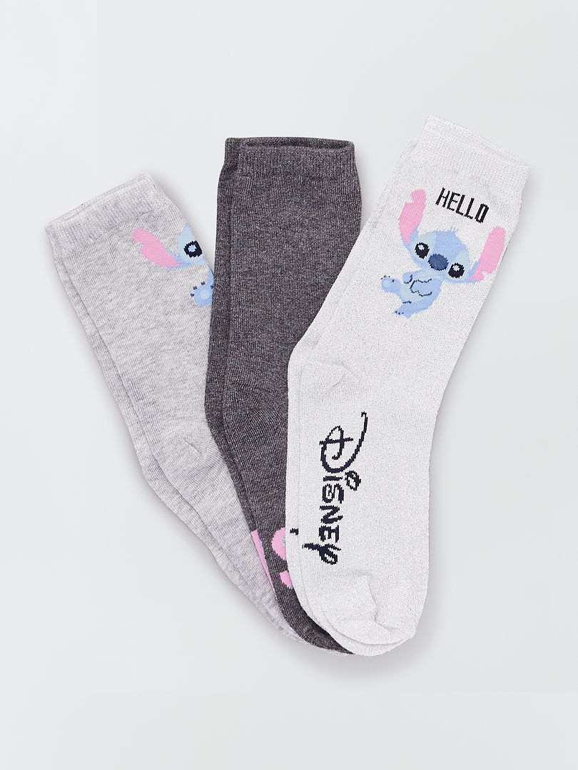 Lot de 3 paires de chaussettes 'Disney14 Jul 2023
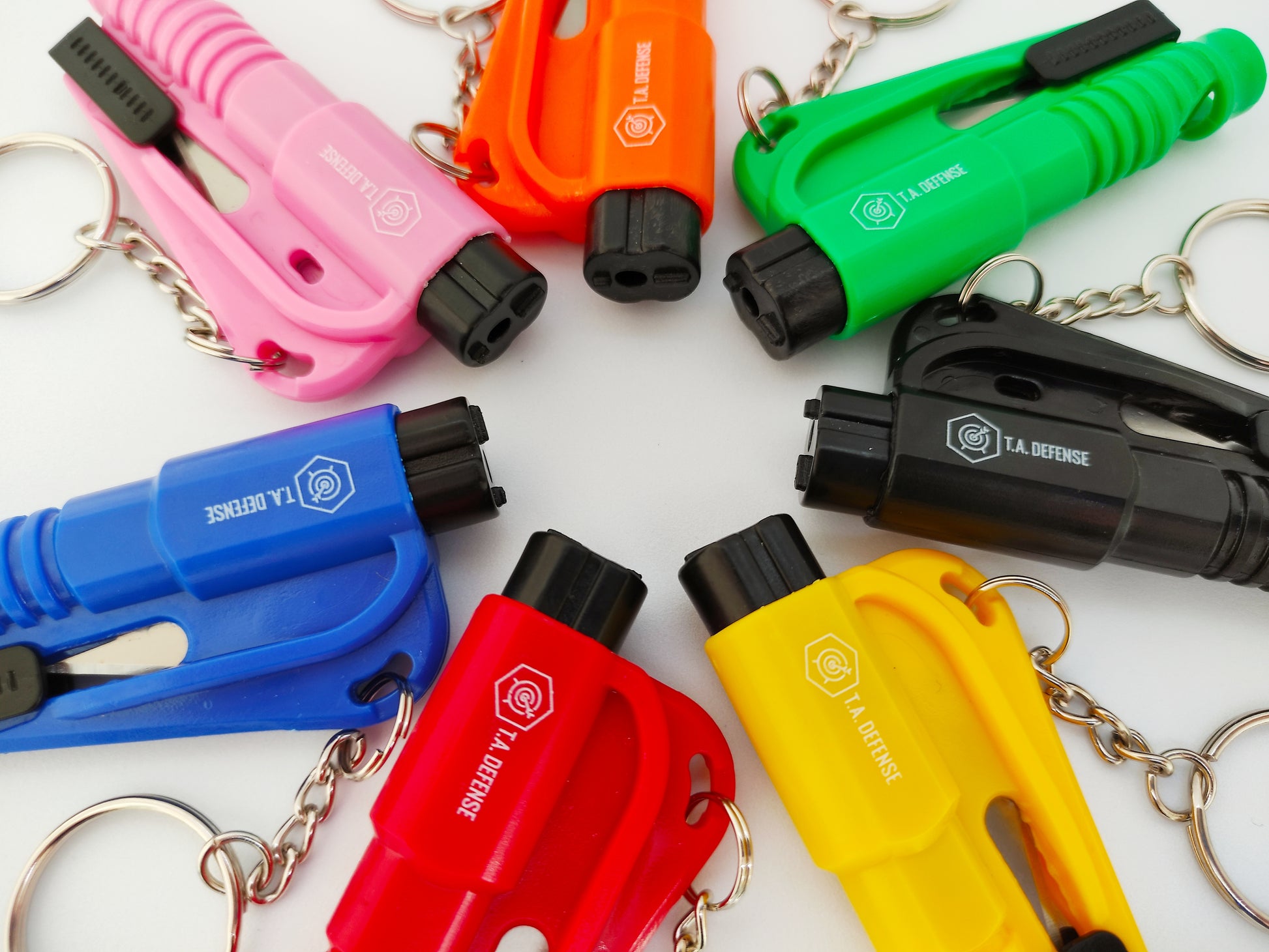 Brise vitre et coupe ceinture, Kiker14 Jul 2023
 Porte papiers voiture bleu pétrole+ étui carte grise made in france simili cuir14 Jul 2023
 Quntis LED USB Lampe de Bureau Écran Ordianteur Anti-lumière Bleue Luminosité/Couleur Réglable, Auto Lumière Mode, Light Bar Moniteur Éclairage Professionnel Barre Lumineuse Protection des Yeux 52cm : : Luminaires et Éclairage14 Jul 2023
 Rowenta Mini Excel Chauffage soufflant céramique 2-en-1, Radiateur14 Jul 2023
 mDesign boite de rangement plastique avec tiroir – bac de rangement plastique bas pour les chaussures – boite empilable pour accessoires, etc. – lot de 2 – transparent : : Cuisine et Maison14 Jul 2023
 Sono portable sur batterie, 15 bluetooth, 1 micro main UHF, effet à leds14 Jul 2023