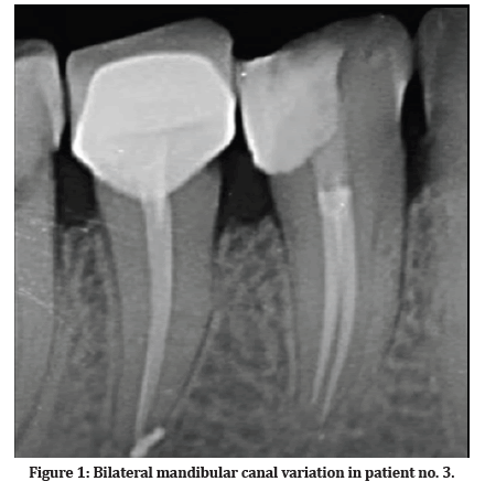
CBCT: 3D image with identification of the left bifid mandibular canal.
Par un écrivain mystérieux
Last updated 23 mai 2024


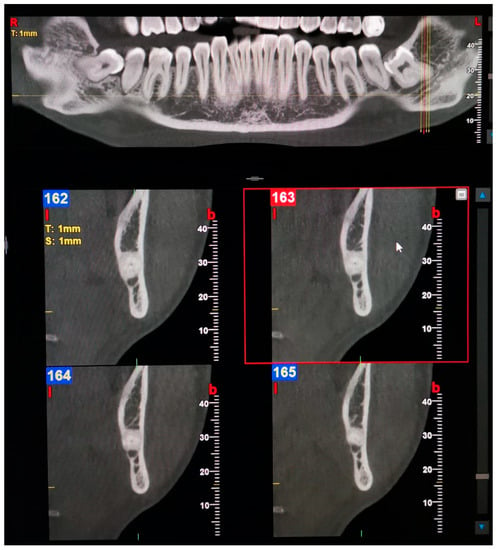
Prevalence of bifid mandibular canal according to gender, type and

JaypeeDigital

Morphology and Variant Anatomy of the Mandibular Canal in a Kenyan

CBCT Anatomical Review of the Mandible

BIR Publications

Directions of mandibular canal displacement in ameloblastoma: A
Oral, Free Full-Text

Figure 7 from Mandibular Canal Enlargement: Clinical and

Morphology and Variant Anatomy of the Mandibular Canal in a Kenyan
Tomographical Determination of Uncommon Mandibular Canal Variations

OPG: left bifid mandibular canal (white arrows). OPG

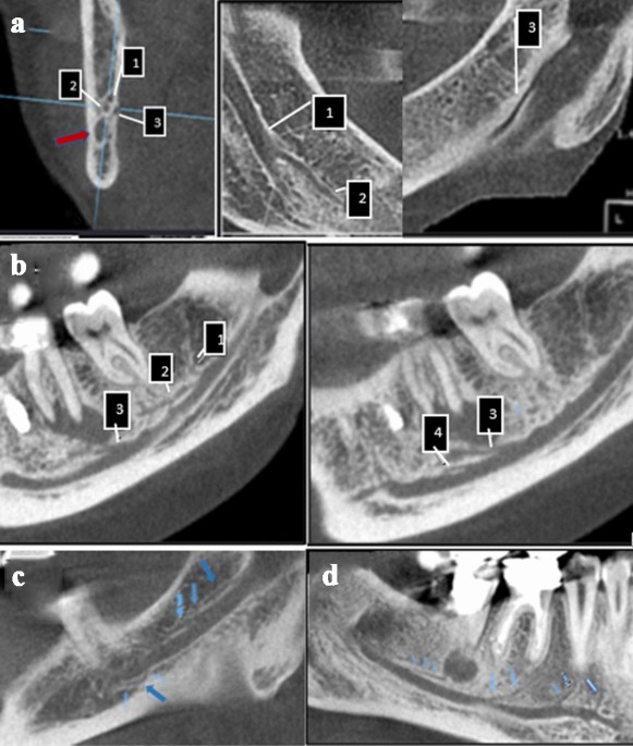
CBCT images of each type of the bifid mandibular canal. A. The
Prevalence of bifid and trifid mandibular canals with unusual
Recommandé pour vous
- SketchUp - How do I create a canal14 Jul 2023
![Male Inguinal Canal - Download Free 3D model by lukaidarch [ad453d5] - Sketchfab](https://media.sketchfab.com/models/ad453d52d93648d886de5b2730f6dd74/thumbnails/4522c2d57f6c45ad95c5c6e4f35bcf70/5e9c4b96c9f74a4fad2b85fc3db22a0f.jpeg) Male Inguinal Canal - Download Free 3D model by lukaidarch [ad453d5] - Sketchfab14 Jul 2023
Male Inguinal Canal - Download Free 3D model by lukaidarch [ad453d5] - Sketchfab14 Jul 2023 Going to Amsterdam? Visit the 3D printed house14 Jul 2023
- canal - - 3D Warehouse14 Jul 2023
 Amsterdam architects plan 3D-printed canal house14 Jul 2023
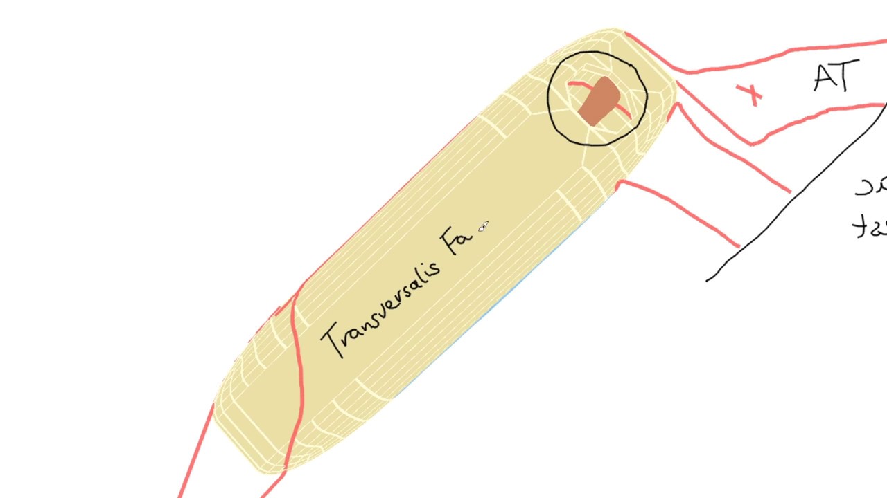 3D Tour of the Inguinal Canal14 Jul 2023
 Hood Canal, WA Custom Map — 3D WOOD MAPS - BELLA MAPS14 Jul 2023
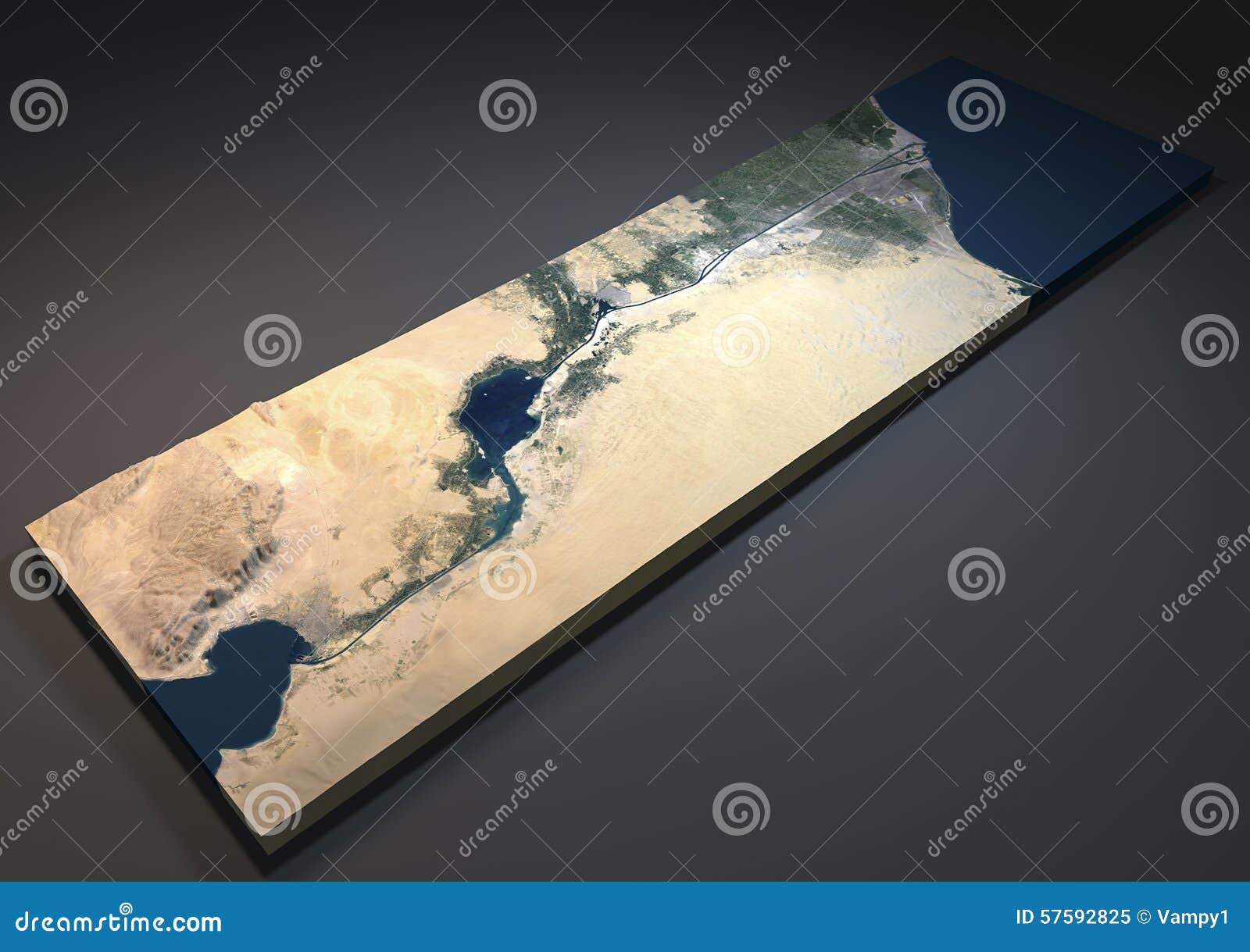 Suez Canal in 3d, Satellite View, Egypt Stock Illustration - Illustration of port, traffic: 5759282514 Jul 2023
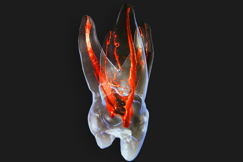 Root Canal - Starlight Dental Clinic14 Jul 2023
 Canal+ cesa las emisiones de su canal 3D en Francia14 Jul 2023
Tu pourrais aussi aimer
 1pcs Evil Bad Banana Man Stuffed Plush Toy Evil Banana Fruit Cute Soft Stuffed Doll for Wedding Gift Toys Valentine's Day Gift14 Jul 2023
 Serpillière ménage à plat NEO 3 NEO 3 ELEPHANT14 Jul 2023
 Super Lance-toiles Spider-Man Marvel Avengers Hobby14 Jul 2023
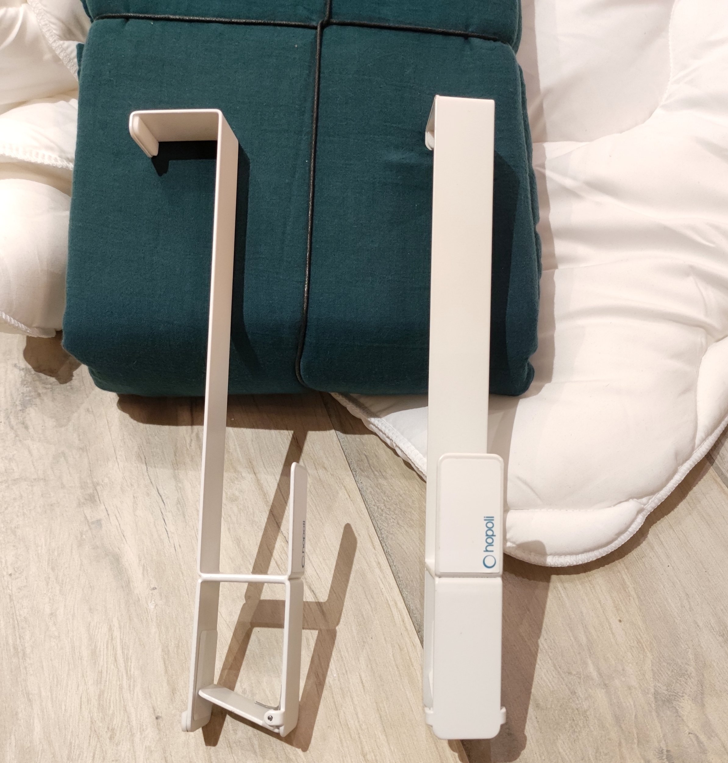 Hopoli, enfile-couette 3-en-1 - IDEES CADEAUX - Autour du Blanc14 Jul 2023
 Circulateur Chauffage Yonos PICO 25-40 180 mm WILO - eco-bricolage14 Jul 2023
 Universal - Magnétophone en direct + microphone à collier Enregistrement numérique Mini magnétophone Enregistrement vidéo DSLR Microphone Mariage en direct - Enregistreur audio numérique - Rue du Commerce14 Jul 2023
 Stanley Vacuum Thermos Bottle Coffee Classic Hammertone 1.1 Q 1 Liter BPA Free14 Jul 2023
 Mousseur à lait SM 3584 SEVERIN14 Jul 2023
 R39 Reflector Tungsten Filament Spotlight Bulb Lamp SES E14 25W at Rs 1379.00, Tungsten Filaments14 Jul 2023
 Organisateur de tiroir en nid d'abeille - Séparateur de tiroir14 Jul 2023